Home
Uncategories
Muscles Of The Chest And Abdomen Labeled / Muscles Of The Thorax Abdomen Anatomy Model Youtube - The muscle striations, are they easily visible on the cat as they are in the dissection book or are they procedure:
Muscles Of The Chest And Abdomen Labeled / Muscles Of The Thorax Abdomen Anatomy Model Youtube - The muscle striations, are they easily visible on the cat as they are in the dissection book or are they procedure:
Muscles Of The Chest And Abdomen Labeled / Muscles Of The Thorax Abdomen Anatomy Model Youtube - The muscle striations, are they easily visible on the cat as they are in the dissection book or are they procedure:. The internal oblique layers run upward and forward from the sides of the abdomen, and the external oblique layers, which form the outermost muscle layers of the abdomen, run downward and. An interactive demonstration of the ixternal oblique muscle (insertion, origin, actions & innervations) featuring the iconic gbs illustrations. Major nerves, vessels and organs are located on the inner surface of the posterior abdominal wall. Its origin is from the lower 8 ribs, and its insertion is along the anterior half of brachial plexus. As the abdominal muscles are hard to support externally, treatment involves rest and pain medication.
The abdominal head of the pectoralis major muscle is one of three origins for the pectoralis major. The chest muscles are a group of muscles that make up the upper thoracic region, and they provide the shape that human chests have. Respiratory muscle training online course: Muscular wall separating the chest and abdomen. Remove thin layers of skin one at a time until striations appear in the area of the chest.
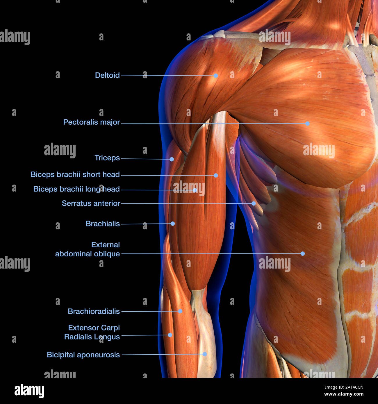
Male Muscle Model from classroom.sdmesa.edu Anterior surface of the sternum, the superior six costal cartilages, and the aponeurosis of the external oblique muscle. The skeletal muscles of the abdomen form part of the abdominal wall, which holds and protects the gastrointestinal system. How to build ab and chest. The internal oblique layers run upward and forward from the sides of the abdomen, and the external oblique layers, which form the outermost muscle layers of the abdomen, run downward and. Muscles of the chest enable us to lift, extend, and rotate our arms, along with playing a part in the process of respiration. Free online quiz muscles of the chest and abdomen labeling. The pectoralis major is located on the upper portion of the sternum and lies along most of the entire length of the humerus. Small muscles running between the ribs, known as the external intercostal muscles, lift the ribs during deep breathing to further expand the chest and lungs and provide even more air to the body.
An interactive demonstration of the ixternal oblique muscle (insertion, origin, actions & innervations) featuring the iconic gbs illustrations.
The external oblique muscle is a broad muscle that runs along the anterolateral abdomen and chest wall. Chest muscles function in respiration while abdominal muscles function in torso movement and in maintenance of balance and posture. As the abdominal muscles are hard to support externally, treatment involves rest and pain medication. Innervation for muscles with chest wall attachments are labeled. Common chest and abdominal injuries. Here is the same image with the chest muscles labeled. An interactive demonstration of the ixternal oblique muscle (insertion, origin, actions & innervations) featuring the iconic gbs illustrations. Fabian identifying the muscles and landmarks of the abdomen and chest. It is the major muscle that the body uses for breathing. Respiratory muscle training online course: There are multiple functions of these chest muscles. Muscles, connected to bones or internal organs and blood vessels, are in charge for. The posterior abdominal wall is found medial to the lateral abdominal walls and is limited.
Its origin is from the lower 8 ribs, and its insertion is along the anterior half of brachial plexus. Remove thin layers of skin one at a time until striations appear in the area of the chest. The muscle striations, are they easily visible on the cat as they are in the dissection book or are they procedure: Here is the same image with the chest muscles labeled. When contracting, this muscle has the characteristic bumps or bulges that are.
Muscles Of The Anterior Thorax And Abdomen Isaiah S Anatomy Website from isaiahzimmermananatomy.weebly.com Chest muscles function in respiration while abdominal muscles function in torso movement and in maintenance of balance and posture. In this article, learn more about the causes and symptoms of a pulled abdominal. Fabian identifying the muscles and landmarks of the abdomen and chest. The abdominal muscles stretch over the abdomen from the chest to the hips, covering the center and sides also. Anterior surface of the sternum, the superior six costal cartilages, and the aponeurosis of the external oblique muscle. Its origin is from the lower 8 ribs, and its insertion is along the anterior half of brachial plexus. Check out this library of free labeling diagrams. Linea alba (white line of connective tissue at midline).
It is the major muscle that the body uses for breathing.
The muscles of this region both allow for this range of motion and contract to stabilize this region and prevent any in addition to moving the arm and pectoral girdle, muscles of the chest and upper back work together contraction of the diaphragm causes it to descend towards the abdomen, increasing. How to build ab and chest. The abdominal head of the pectoralis major muscle is one of three origins for the pectoralis major. Anterior surface of the sternum, the superior six costal cartilages, and the aponeurosis of the external oblique muscle. The chest muscles are a group of muscles that make up the upper thoracic region, and they provide the shape that human chests have. Here is the same image with the chest muscles labeled. Respiratory muscle training strengthen the function of the respiratory muscles to improve your patient's overall performance powered by physiopedia start. The muscle striations, are they easily visible on the cat as they are in the dissection book or are they procedure: Innervation for muscles with chest wall attachments are labeled. The posterior abdominal wall is found medial to the lateral abdominal walls and is limited. Fabian identifying the muscles and landmarks of the abdomen and chest. The upper part of the trunk is the chest and the lower one is the abdomen. For some smaller muscle observations, larger.
The skeletal muscles of the abdomen form part of the abdominal wall, which holds and protects the gastrointestinal system. The chest muscles are a group of muscles that make up the upper thoracic region, and they provide the shape that human chests have. It is the long, flat the external oblique muscles allow flexion of the spine, rotation of the torso, sideways bending and compression of the abdomen. Labeling muscles (chest and abdomen). Remove as much adipose tissue and fascia as you can so that the fibers of the muscles can be seen.
Labeled Anatomy Chart Of Male Biceps And Chest Muscle On Black Background Stock Photo Alamy from c8.alamy.com There are multiple functions of these chest muscles. Common chest and abdominal injuries. Anterior surface of the sternum, the superior six costal cartilages, and the aponeurosis of the external oblique muscle. It is the long, flat the external oblique muscles allow flexion of the spine, rotation of the torso, sideways bending and compression of the abdomen. Well, imagine you're standing up straight, or you can just go the chest is separated from the abdomen by the diaphragm, a large smooth muscle that enables us to breathe by changing the air pressure in the lungs. The primary function is certainly to provide support to the skeletal system and to facilitate its movements. It is the major muscle that the body uses for breathing. Muscles of the chest enable us to lift, extend, and rotate our arms, along with playing a part in the process of respiration.
Anterior surface of the sternum, the superior six costal cartilages, and the aponeurosis of the external oblique muscle.
How to build ab and chest. Extend your arms (and the band) fully in front of your chest, then. Remove as much adipose tissue and fascia as you can so that the fibers of the muscles can be seen. The primary function is certainly to provide support to the skeletal system and to facilitate its movements. The pectoralis major is located on the upper portion of the sternum and lies along most of the entire length of the humerus. Muscles of the chest enable us to lift, extend, and rotate our arms, along with playing a part in the process of respiration. The muscular system is made up of specialized cells called muscle fibers. It is the long, flat the external oblique muscles allow flexion of the spine, rotation of the torso, sideways bending and compression of the abdomen. Respiratory muscle training strengthen the function of the respiratory muscles to improve your patient's overall performance powered by physiopedia start. Free online quiz muscles of the chest and abdomen labeling. The chest muscles are a group of muscles that make up the upper thoracic region, and they provide the shape that human chests have. The skeletal muscles of the abdomen form part of the abdominal wall, which holds and protects the gastrointestinal system. The posterior abdominal wall is found medial to the lateral abdominal walls and is limited.
Common chest and abdominal injuries muscles of the chest abdomen. Muscular wall separating the chest and abdomen.


0 Comments:
Posting Komentar